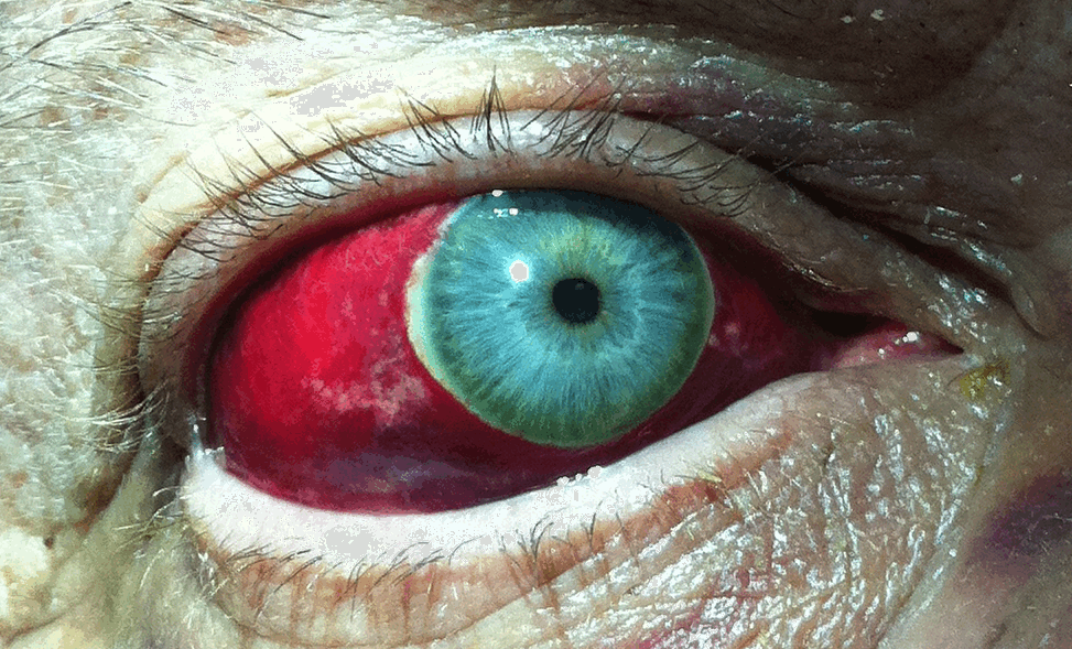
Occasional Yealy Sub Eye Bleeds
Hyphema, or blood in the anterior chamber of the eye, is a common condition among cats. However, hyphema is a clinical sign and not a specific disease in itself. Symptoms and Types. The symptoms of hyphema are dependent on the extent of bleeding, whether vision has been impaired, and whether your cat has other systemic diseases. Sub-conjunctival haemorrhage. A sub-conjunctival haemorrhage is caused by a bleeding blood vessel under the conjunctiva. Patients will often present after being told they have a red eye and may not have noticed any symptoms. They usually have no cause but are more common after coughing or vomiting excessively. Can also be caused by mild trauma.
Retinal hemorrhages are often hallmarks of many ocular and/or systemic diseases. Thus, finding them in asymptomatic patients during comprehensive eye exams may require further evaluation to determine the principle cause. It is crucial to identify and classify various types of hemorrhages because optometric management is influenced by the underlying etiology. The following are the most common categories of retinal hemorrhages and their associated diseases.
1. Subhyaloid & pre-retinal hemorrhages = “D or boat-shaped retinal heme”
Occasional Yearly Sub Eye Bleeds Symptoms
- Location:
- Subhyaloid heme – between posterior vitreous base & internal limiting membrane.
- Pre-retinal heme – posterior to internal limiting membrane & anterior to nerve fiber layer.
- Possible etiologies:
- Retinal neovascularization
- Posterior vitreous detachment & retinal breaks (result of tearing the major retinal vessels)
- Terson’s syndrome (vitreous heme + subarachnoid heme)
- Retinal trauma
- Valsalva retinopathy
2. Flame-shape hemorrhages = “Feathered” or linear retina heme
- Location: within nerve fiber layer (resolve around six weeks)
- Possible etiologies:
- Hypertensive retinopathy
- Papilledema
- Anterior ischemic optic neuropathy
3. Dot-and-blot hemorrhages = “Round” retinal heme
- Location: inner nuclear & outer plexiform layers (resolve time is longer than flame-shaped hemes)
- Possible etiologies:
- Idiopathic juxtafoveal retinal telangiectasis
- Ocular ischemic syndrome
4. Subretinal & subretinal pigment epithelium (RPE) hemorrhages = dark color retinal heme
- Location: beneath neurosensory retina (resolve very slowly)
- Subretinal heme – heme in space between neurosensory retina & retinal pigment epithelium (have amorphous shape due to absence of firm attachments between neurosensory retina & RPE).
- Sub-RPE heme – heme located between RPE and Bruch’s membrane (have well-defined borders because of tight cell junctions between RPE cells).
- Possible etiologies:
- Choroidal neovascular membrane formation
- Neurosensory or RPE detachments
- Wet age-related macular degeneration
- Choroidal tumors
- Trauma

Occasional Yealy Sub Eye Bleeds Causes
Reference:
Occasional Yearly Sub Eye Bleeds Twice
- Shechtman, D., Kabat, A. “The Many Faces of a Retinal Hemorrhage.” Optometric Management. [Retrieved August 5, 2013] http://www.optometricmanagement.com/articleviewer.aspx?articleid=101343